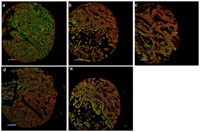
Figure 3. AQUA quantitative image analysis.
DAPI counterstain (blue) was used to identify nuclei, pan-cadherin antibody (rabbit) or a combination of pan-cadherin and pan-cytokeratin antibodies (mouse) to identify infiltrating tumour cells (green) and Cy-5-tyramide for detection of following target proteins (red) (a. E-cadherin, b. SNAIL, c. SLUG, d.WT1 and e. phospho-β-catenin).

