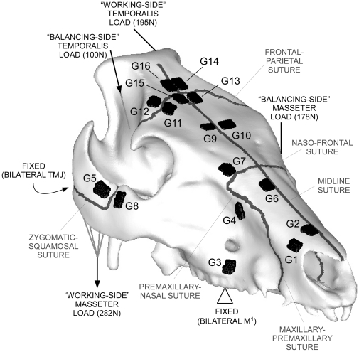
Figure 1. CT reconstruction of the pig skull for FE modelling.
CT reconstruction of the pig skull, showing the locations and magnitudes of loading and constraints used in the FE model, as well as the positions of the strain gauges and sutures. TMJ = temporomandibular joint, M1 = 1st molar tooth, G = gauge.

