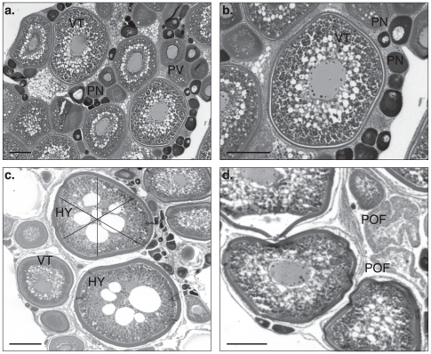
Figure 2. Histology of Spawning Black Marlin Ovary.
A) Perinucleolar oocytes (PN), previtellogenic oocytes (PV) and vitellogenic oocytes (VT) displaying incremental development within these stages. B) Vitellogenic oocyte displaying a prominent nucleus with numerous peripheral nucleoli surrounded by yolk granules. C) early stage hydrated oocytes (HY) with large yolk globules and the 3 axes used to measure distorted oocytes. D) Post ovulatory follicles (POF) alongside advanced vitellogenic oocytes. Scale bar 200 µm.

