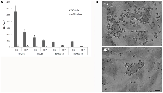
Figure 1. Adhesion of asexual stages of ItG and 3D7 on stimulated and non stimulated HDMEC, HUVEC and HBMEC endothelial cell lines.
A) Data shown are the mean number of iRBC per mm2 ± S.E. of 3 to 5 biological replicates. Static assays were carried out as described in the Material and Methods section. B) Giemsa-stained infected erythrocytes bound to TNF-alpha activated endothelial cells (HDMEC). Scale bar: 25 µm.

