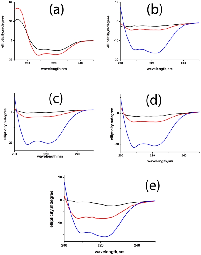
Figure 7. Effect of TFE on the structure of WT and cysteine mutants of Viperin monitored by Far-UV CD.
Far UV-CD spectra of (a) the WT (b) the triple cysteine mutant (c) C83A mutant (d) C87A mutant and (e), C90A mutant of Viperin in the presence (red) and absence of 50% TFE (black). Far UV-CD spectrum of WT Viperin (blue) has also been shown (7(b)-7(e)). All these experiments have been carried out in 20 mM sodium phosphate buffer at pH 7.5.

