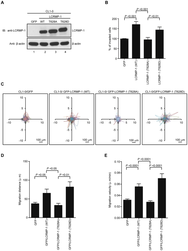
Figure 2. Phosphorylation of LCRMP-1 at Thr-628 is critical for cancer invasion and migration.
(A) Protein expression levels of exogenous untagged LCRMP-1 (WT), LCRMP-1 (T628A), and LCRMP-1 (T628D) are confirmed by immunoblotting. After 48 hours post lentivirus infection, CL1-0 cells were stably expressing wild-type and mutant LCRMP-1. Lentivirus expressing GFP served as control. Cell lysates were harvested, following by assessing with immunoblotting using anti-LCRMP-1 antibody and anti-β-actin antibodies. (B) Nonphosphorylated LCRMP-1 (T628A) mutant lowers activity of cell invasion. The invasive capacity of these cells was determined with the modified Boyden chambers invasion assay in vitro. Percentage of invasive ability was normalized to GFP control. Data were shown as means ± SEM for three-independent experiments (n = 3). (C) Nonphosphorylated LCRMP-1 (T628A) mutant greatly suppressed cell migration tracks. Tract plots showed CL1-0 cells expressing GFP, GFP-LCRMP-1 (WT), GFP-LCRMP-1 (T628A), GFP-LCRMP-1 (T628D), respectively. Moving tracks of at least 10 representative cells at the start point all set to ‘0,0’ over a 20-hour period (different lines) showed the representative motility of cells. Scale bar, 100 µm. (D, E) Total migration distance (D) and cell migration velocity (E) were quantified from cell tracking assay for 20 hours. Data were presented as means ± SEM.

