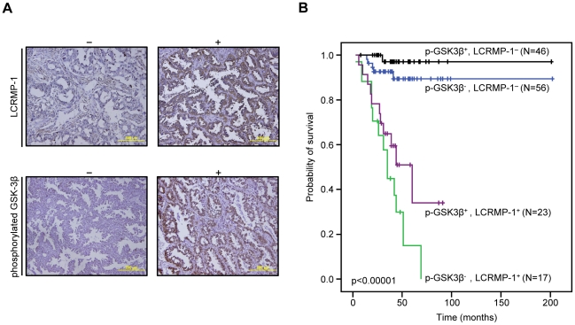
Figure 5. Kaplan-Meier survival plots for NSCLC patients grouped by phosphorylated GSK3β and LCRMP-1 protein expression levels.
(A) Typical protein expression patterns of phosphorylated GSK3β and LCRMP-1 were detected by immunohistochemistry using anti-phospho-GSK3β (Ser9) and anti-LCRMP-1 antibodies (C2) in serial dissections of primary tumor specimens from 142 NSCLC patients who underwent surgical resections. Results are shown + and − denotes tumors with and without over-expression with indicated protein respectively. Scale bars, 100 µm. p-GSK3β was represented to phosphorylated GSK3β. (B) Kaplan–Meier analysis of overall survival for 142 NSCLC patients with p-GSK3β−-LCRMP-1−, p-GSK3β−-LCRMP-1+, p-GSK3β+-LCRMP-1−, and p-GSK3β+-LCRMP-1+. P values were performed by 2-sided log-rank tests.

