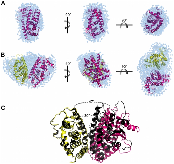
Figure 4. SAXS models for LBD proteins construction.
Three orthogonal views of the SAXS ab initio models for A) hPPARγ LBD, obtained by Gasbor (shaded spheres), superposed to the hPPARγ LBD monomeric part of the high-resolution model PDB id 1FM6 (cartoon) and B) hPPARγ/hRXRα LBD heterodimer, obtained by Gasbor (shaded spheres), superposed to the rigid body model from PDB id 1FM6 (cartoon). C) Superposition of the rigid body model with the crystallographic structure (PDB id 1FM6) showing the opening angle imposed on the rigid body model being larger than the crystallographic structure. hPPARγ LBD (pink), hRXRα LBD (yellow), crystallographic structure of heterodimer (black) and DAM (blue).

