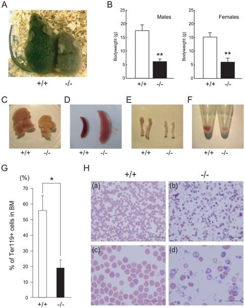
Figure 2. Growth retardation and peripheral blood morphology in postnatal CALM-deficient mice.
(A) Wild-type (+/+) (left) and CALM-deficient (−/−) (right) mice at 3 weeks of age. (B) Average weight of wild-type (+/+) and CALM-deficient (−/−) mice at postnatal day 28. Data shown represent CALM-deficient males (n = 3), CALM-deficient females (n = 4), wild-type males (n = 36), and wild-type females (n = 35). **, P<0.01 vs control. Liver (C), spleen (D) and tibia (E) from wild-type (+/+) or CALM-deficient (−/−) mice at 3 weeks of age. (F) Bone marrow cells were collected by centrifugation. Note that the pellet from CALM-deficient (−/−) mice is small and pale in color. (G) Quantification of TER119-positive bone marrow cells (data represent means±SD; n = 5). *, P<0.05 vs. control. (H) Peripheral blood smears from wild-type (+/+) littermates (left panels, a and c) or CALM-deficient (−/−) mice (right panels, b and d) at 3 weeks of age. Magnification: upper panels, 400×; lower panels, 1000×. Scale bar, 50 µm (upper panel) and 20 µm (lower panel).

