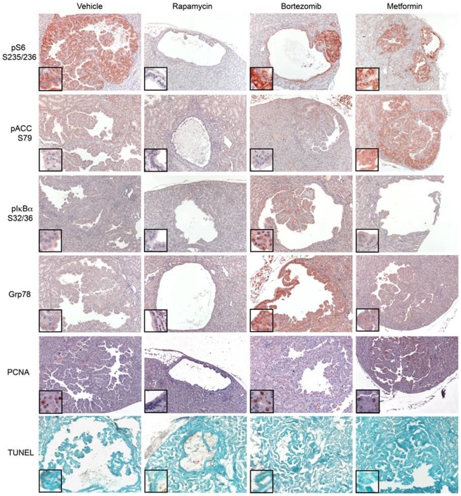
Figure 4. Immunohistochemistry (IHC) analysis of therapeutic effects.
Tsc2+/− A/J strain mice of ages 9–10 months were treated with drugs for 1 week, and kidneys prepared for histology. Sections were prepared and stained using pS6-S235/236 (red), pACC-S79 (red), pIκBα-S32/34 (red) and GRP78 (red) antibodies. The bottom panel shows apoptosis assessed by TUNEL method. Representative sections are shown. All images shown are taken at 100× magnification. Insets show portions of the tumor at higher magnification (400×).

