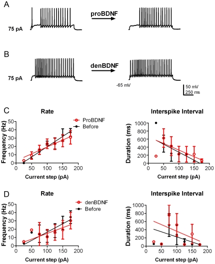
Figure 2. Neither proBDNF nor denatured BDNF increase mitral cell excitability.
(A–B) Representative action potential trains in mitral cells in response to 75 pA current injections of 1000 ms duration before (left) and approximately 10 min after (right) bath application of either 10 ng/ml proBDNF (A) or 10 ng/ml denatured BDNF (B, denBDNF). The resting membrane potential was held near −65 mV. (C–D) Line graphs of the mean (± s.e.m.) spike frequency (Hz) and of the mean (± s.e.m.) ISI at each current injection as recorded in A and B before (closed circles; black) and after (open circles; red) proBDNF (C) or denatured BDNF (D). Data were fit with linear regressions to facilitate visualization. Neither treatment had a significant effect upon either rate or ISI; two-way ANOVA (N = 4; p≥0.05).

