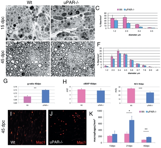
Figure 3. Sciatic nerve regeneration after injury.
(A–B, D–E) semithin section and (C, F) fiber type distribution from sciatic nerve of Wt and uPAR null mice at 15 and 45 dpc. At both 15 and 45 dpc we observed reduced number of regenerating fibers. (G) g-ratio was significantly increased in uPAR−/− regenerating fibers at 45 dpc (n. 20000; p = 0.01). (H) Neurophysiological analysis showing similar values of cMAP between Wt and uPAR−/− mice, whereas NCV was significantly reduced in uPAR−/− mice (n. 8; p = 0.001). (I–J) Staining for Mac-1/CD11b (Mac1) in Wt (I) and uPAR null (J) sciatic nerve 45dpc. (K) Quantification of number of macrophages observed in sciatic nerve 15, 21 and 45dpc; differences were significant at 21 and 45 dpc (*p = 0.04; **p = 0.008). Bar = 10 µm in A, B, D, E, I and J.

