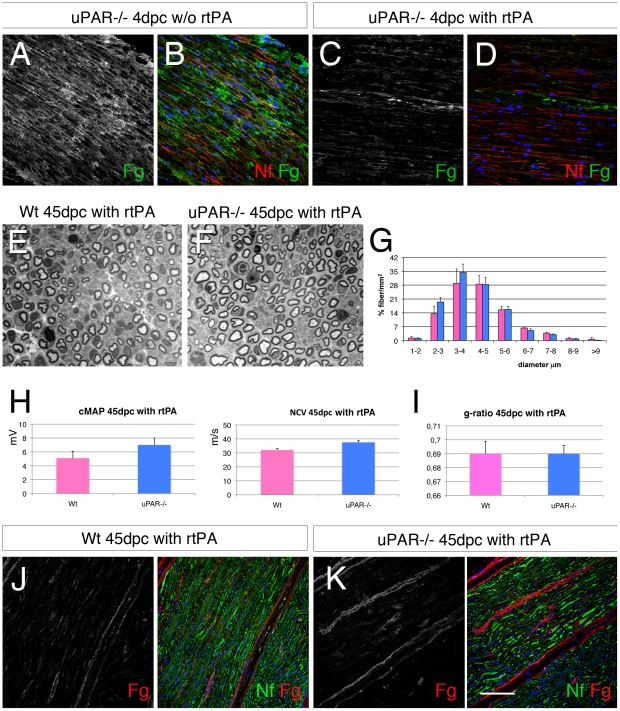
Figure 6. Exogenous recombinant tPA rescues abnormal myelination in uPAR null nerves after injury.
(A–D) Sciatic nerve cryosections from Wt and uPAR null mice stained for fibrin 4 dpc without or after exogenous treatment with recombinant tPA (5 mg/100 g). After treatment fibrin staining is mostly abolished (compare C and A). (E–F) semithin sections from sciatic nerve 45 dpc of Wt and uPAR null mice after treatment with recombinant tPA (5 mg/100 g , 2 times per week for 3 weeks), and (G) fiber type distribution. Number and fiber type distribution was similar in the two groups. (H) Neurophysiology analysis showing similar values of cMAP and NCV between Wt and uPAR−/− mice treated with rtPA at 45 dpc. (I) g-ratio did not show differences in myelin thickness between Wt and uPAR−/− nerves (n. 15000). (J–K) immunofluorescence staining for fibrin in sciatic nerves at 45 dpc from mice treated with rtPA, showing low expression of fibrin. Fg = fibrin; Nf = neurofilaments. Bar = 50 µm in A–D and J–K; 20 µm in E–F.

