
Fig. 1.
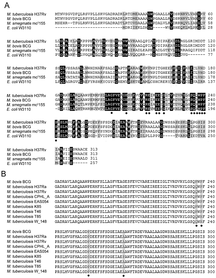
Multiple alignments of NudC from different origins. Sequences aligned using clustal w.
A. Alignments of NudC from E. coli W3110 (AP_003822), M. tuberculosis H37Rv (NP_217715), M. smegmatis mc2155 (YP_886312) and M. bovis BCG (YP_979308). Conserved residues (Nudix box and NudC characteristic sequences) in NudC are highlighted (•).
B. Alignments of NudC from different M. tuberculosis clinical isolates and M. bovis BCG. Only regions containing differences are highlighted (grey and •).