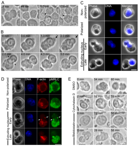
Fig. 1.
Enucleation is initiated through establishment of cell polarization, followed by contractions of asymmetrically localized cytoplasmic actomysoin. (A) Time-lapse images of late erythroblasts undergoing cell polarization. White dots depict the nucleus. Time elapsed in minutes after completion of the final mitotic division of the erythroid progenitor cell is shown. (B) Time-lapse images of late erythroblasts undergoing enucleation. (C) Late erythroblasts were fixed and stained for DNA (blue). The populations of late erythroblasts were classified into non-polarized cells, polarized cells that show nuclear displacement, early phase of nuclear extrusion and late phase of nuclear extrusion. (D) Immunofluorescence of late erythroblasts at different stages of enucleation. Single confocal images of F-actin (red), active myosin II (pMRLC; green) and DNA (blue) are shown. Note that both F-actin and active myosin II become mostly restricted to the opposite side of the cytoplasm after cells become polarized. A small fraction of both F-actin and active myosin (arrows) is found accumulated in the neck region of the bleb-like protrusion. (E) DMSO, cytochalasin D or blebbistatin was applied to cells at 36 hours and then polarized late erythroblasts were identified for time-lapse recording. Scale bars: 5 μm.

