Fig. 2.
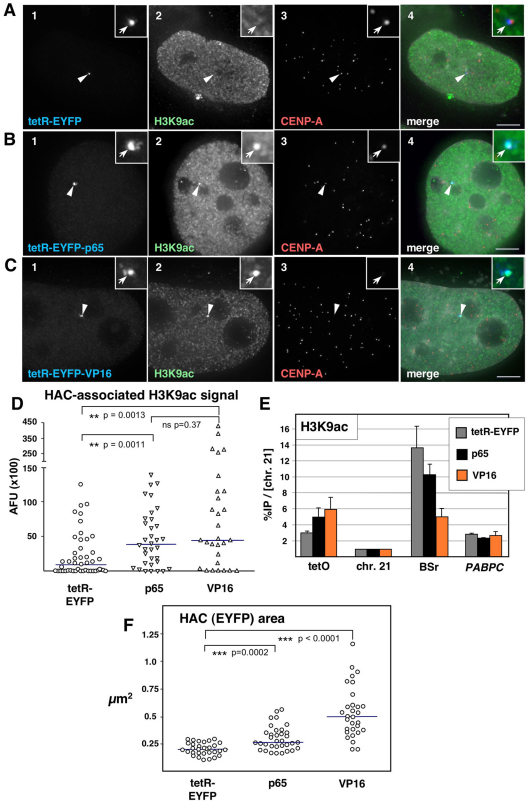
Tethering of VP16 and p65 mediates HAC centromere hyper-acetylation. (A–C) Immunofluorescence (IF) analysis of 1C7 cells 2 days after transfection with the indicated tetR fusion constructs. Cells were co-stained with antibodies against acetylated H3K9 (H3K9ac, green, panel 2) and CENP-A (red, panel 3). Arrowheads depict the HAC, as determined by the EYFP signal (blue, panel 1). Merged images (panel 4) represent the overlay of EYFP signals with antibody and 4′,6-diamidino-2-phenylindole (DAPI) staining (light grey). Scale bars: 5 μm. (D) Fluorescence signals of HAC-associated H3K9ac staining in individual cells transfected as in A–C were quantified and plotted as arbitrary fluorescence units (AFU). Solid lines indicate the median. (E) ChIP analysis as in Fig. 1C of 1C7 cells transfected with tetR–EYFP, tetR–EYFP–p65 (p65) or tetR–EYFP–VP16 (VP16), using an antibody against H3K9ac. Values are normalized to the chromosome 21 centromere locus and represent the mean and s.e.m. of three independent experiments. (F) Quantification of HAC decondensation in cells transfected as in A–C. The HAC-bound EYFP area was measured in maximum-intensity projections of cells expressing the indicated fusion constructs. Sold lines indicate the median.

