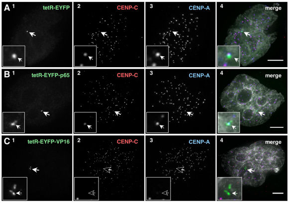
Fig. 5.
Tethered VP16, but not p65, removes CENP-C from the HAC kinetochore. (A–C) Cells expressing the indicated tetR fusion proteins were fixed and stained 2 days after transfection. tetR–EYFP fusion protein (green, panel 1), CENP-C (red, panel 2) and CENP-A (blue, panel 3). Merged images (panel 4) represent the overlay of EYFP signal, antibody and DAPI staining (light grey). Arrows depict the HAC. Scale bars: 5 μm.

