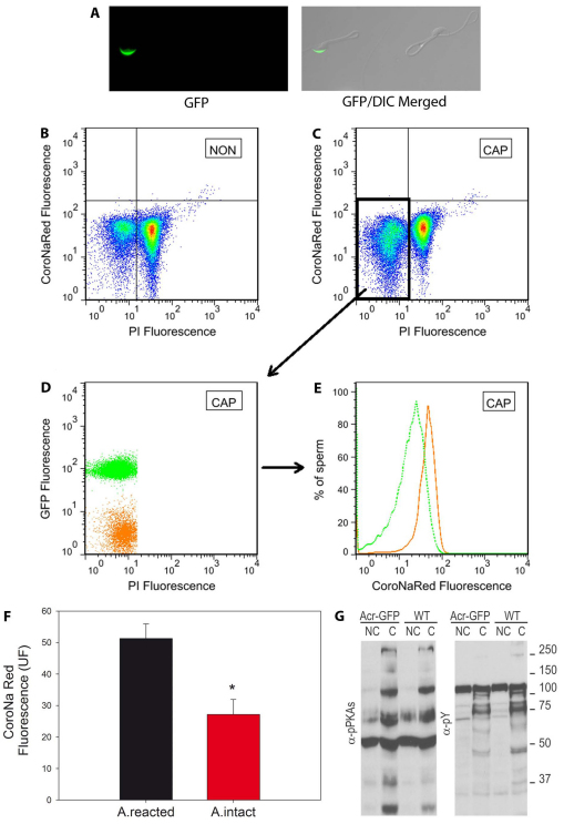
Fig. 2.
[Na+]i decreases only in acrosomal intact sperm. (A) Sperm from the cauda epididymides of Acr-GFP CF1 mice, with GFP in their acrosomes, were incubated in non-capacitating medium. GFP fluorescence (left) and the merged DIC and GFP images (right) of two representative reacted and not-reacted sperm are shown. (B,C) These sperm were loaded with CoroNa Red for 30 minutes and then incubated in the same medium or in medium that supported capacitation. Before flow cytometry, PI was added to the sperm suspension. Two-dimensional CoroNa Red versus PI fluorescence dot plots of Acr-GFP sperm were used to analyze NON (B) and CAP (C) live and dead sperm as described in Fig. 1. (D) The spontaneous acrosome reaction in the capacitated live-sperm population was further analyzed using two-dimensional GFP versus PI dot plots in which two clear populations of cells containing either high GFP (in green), with intact acrosomes, or low GFP (in orange), without acrosomes, can be distinguished. (E) These populations were then analyzed and histograms of their individual [Na+]i CoroNa Red fluorescence constructed. [Na+]i was decreased only in the population with intact acrosomes. (F) Average values of CoroNa Red fluorescence in CAP live-sperm populations. Values are means ± s.e.m., n=5 (*statistically significant, P=0.019, paired t-test). (G) Phosphorylation of PKA substrates and tyrosine phosphorylation in wild-type and Acr-GFP sperm was evaluated using western blotting with anti-PKAS-P (left panel) and anti-Y-P (right panel) antibodies.

