Fig. 5.
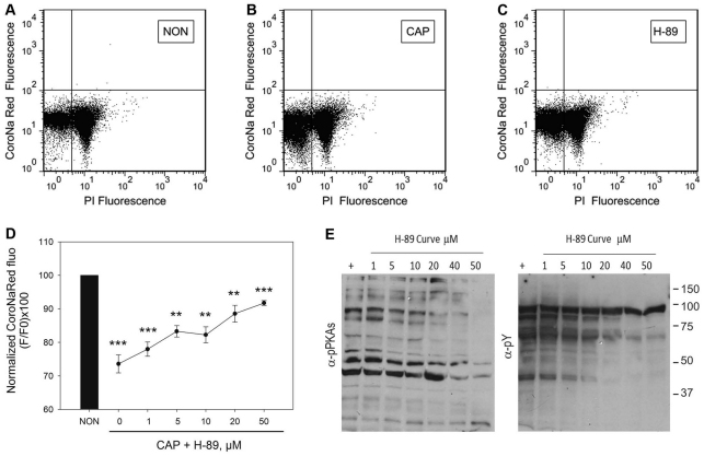
PKA inhibition by H-89 blocks the capacitation-associated decrease in [Na+]i. (A–D) Sperm from the cauda epididymus were placed in medium that did not support capacitation, which was loaded with CoroNa Red for 30 minutes, washed and incubated in the same medium (NON; A) or in medium that supported capacitation (CAP; B) in the absence or in the presence of increasing concentrations of H-89 (0, 1, 5, 10, 20 and 50 μM; C and D). (A–C) Two-dimensional dot plots of PI versus CoroNa Red fluorescence of sperm incubated under non-capacitating (NON) or under capacitating (CAP) conditions in the absence or in the presence of 50 μM H-89. (D) CoroNa Red fluorescence. Values are means ± s.e.m. from four independent experiments (**P≤0.01; ***P≤0.001). (E) Western blots, using anti-PKAS-P (left panel) and anti-Y-P (right panel) of sperm extracts incubated in the same media as the ones used for the flow cytometry.

