Fig. 1.
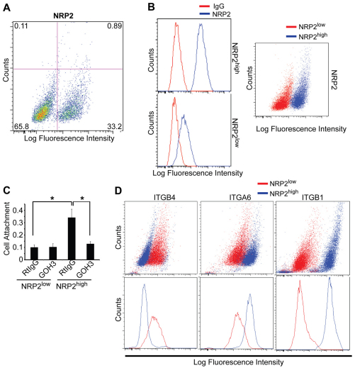
Characterization of NRP2high and NRP2low populations of epithelial cells isolated from human breast tumors. (A) Epithelial cells (EpCAM+) were isolated from breast tumors and analyzed for NRP2 expression by flow cytometry. Approximately 33% of cells express high levels of NRP2. (B) Epithelial cells isolated from four different breast tumors were sorted into NRP2high and NRP2low populations. These populations were stained with either an NRP2 antibody or goat IgG to confirm the relative expression of NRP2. Histograms in the left panels show NRP2 expression relative to control IgG in each population; pseudocolored dot plot in the right panel show NRP2 expression in the NRP2high and NRP2low cell populations. (C) NRP2high and NRP2low populations were incubated with either an α6 antibody (GoH3) or rat IgG for 1 hour, and assayed for adhesion to laminin. NRP2high cells adhere much more avidly to laminin and this adhesion is blocked by GoH3. (D) The relative expression of α6, β1 and β4 integrins in the NRP2high and NRP2low populations was quantified by flow cytometry. The NRP2high population expressed relatively high levels of α6 and β1 integrins, but low levels of the β4 integrin subunit.

