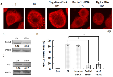
Fig. 5.
P. aeruginosa infection induced autophagy through a classical pathway. (A) MH-S cells were transfected with LC3-RFP plasmids for 24 hours and then infected with PAO1 for 1 hour. Before infection, the cells were also treated with negative control siRNA, beclin-1 siRNA and Atg7 siRNA. Confocal images show LC3 puncta induction. (B) Western blotting of beclin-1. (C) Western blotting of Atg7. GAPDH was probed as a loading control in B and C. (D) Puncta numbers in each cell was determined and cells with more than 10 punctae were considered as LC3-RFP puncta cells. The data are representative of 100 cells (one-way ANOVA; Tukey's post-hoc test, *P<0.05). Data are representative of three experiments with similar results.

