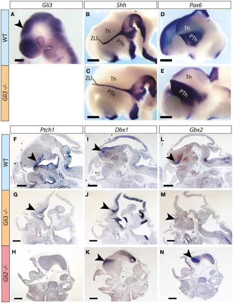
Figure 1.
Thalamic regionalization in the Gli3Xt/Xt mutant. (A–E) Whole mount in situ detection of markers in E9.5 (A) and E12.5 (B–E) mouse embryos, markers, and genotypes as indicated. Rostral to the left. Arrowhead in (A) points to the thalamic primordium. (F–N) In situ detection of marker expression on sections of E12.5 mouse embryos, markers, and genotypes as indicated. Arrowheads point to diencephalic expression. PTh, prethalamus; Th, thalamus; ZLI, zona limitans interthalamica. The brown precipitate in (F,I,L) corresponds to background staining. Scale bar in (A), 200 μm; in (B–N), 500 μm.

