Figure 2.
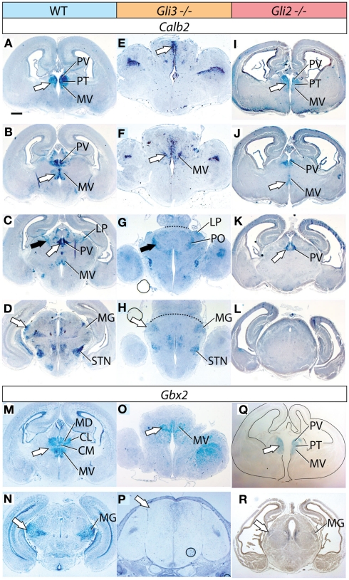
Thalamic differentiation in the Gli3Xt/Xt and Gli2 mutants (1). In situ detection of Calb2 (A–L) and Gbx2 (M–R) expression on transverse sections of wild type (A–D,M,N), Gli3Xt/Xt (E–H,O,P), and Gli2zfd/zfd (I–L,Q,R) E18.5 mouse brains. Comparable rostro-caudal thalamic levels are represented side-by-side. White and black arrowheads point at comparable structures across genotypes. The dotted line in (G) and (H) delimits the mutant thalamus from more caudal structures. The outline of the section has been delineated in (Q). Scale bar, 500 μm.

