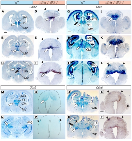
Figure 5.
Thalamic differentiation in the double n-Shh/Gli3 mutant. In situ detection of Calb2 (A–F), Lhx2 (G–L), Gbx2 (M–P), and Cdh6 (Q–T) expression on transverse sections of wild type (A–C,G–I,M,N,Q,R) and n-Shh/Gli3 (D–F,J–L,O,P,S,T) E18.5 mouse brains. Comparable rostro-caudal thalamic levels are represented side-by-side. White arrows and black arrows point at comparable structures across genotypes. The outline of the section has been delineated in (O,P). Scale bar, 500 μm.

