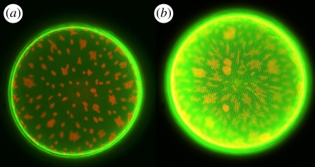
Figure 8.
Comparison of the fluorescence of living diatom cells (Coscinodiscus wailesii) cultivated in Rh 19 concentration of (a) 1 µM and (b) 10 µM. Pictures taken with identical microscope and dual-channel camera settings. Besides the fluorescence of the stained frustule, the autofluorescence of the chloroplasts can be seen. Morphological changes of the chloroplasts are caused by the prolonged exposure to intense light during the experiment (cf. [30]). Valve diameter is approximately 220 µM. (Online version in colour.)

