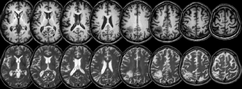
Figure 1.
Axial MRI slices from the patient with neurological damage are shown in neurological convention (with the left side of each slice corresponding to the left side of the brain). Both the T1 (top row) and T2 (bottom row) images have been normalized to MNI stereotaxic space, with slices corresponding to Z = 8, 16, 24, 32, 40, 50, 60 mm.

