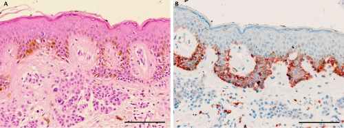
Figure 1.
Representative staining results in a case of compound nevus. A) H&E staining showing a proliferation of junctional and dermal melanocytes with symmetrical pattern and no evidence of cytological dysplasia; B) C-Kit immunohistochemistry showing strong (membranous and cytoplasmatic) expression in the junctional component of compound nevus and weak expression in the dermal component with gradual decrease to the bottom of the lesion. In the reticular dermis frequent c-Kit positive mastcell. Scale bars: 100 μm.

