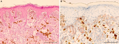
Figure 4.
Representative staining in a case of Spitz nevus. A) H&E staining showing a proliferation of spindle and epitelioid melanocytes with symmetrical pattern and no evidence of cytological dysplasia; B) prominent c-Kit immunohistochemical staining at dermoepidermal junction and progressive decrease positivity in the dermis. Scale bars: 200 µm.

