Fig. 5.
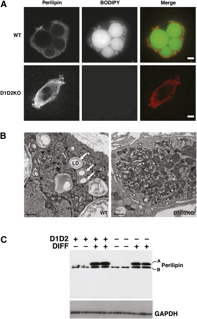
D1D2KO adipocytes express perilipin but lack LDs and accumulate membranous aggregates. A: BODIPY493/503 (BODIPY, green) and perilipin (red) staining of WT and D1D2KO differentiated cells. Scale bar, 5 μm. B: WT and D1D2KO adipocytes visualized by electron microscopy. Lipid droplets are indicated by arrows. Mitochondria are indicated by black arrowheads. A large membranous aggregate is visible (outlined by white arrowheads) in the D1D2KO adipocyte. Aggregates were visible in all D1D2KO adipocytes. Scale bar, 1 μm. C: Western blot of perilipin in undifferentiated (DIFF–) and differentiated (DIFF+) WT (D1D2+) and D1D2KO (D1D2–) cells. A and B indicate the different isoforms of perilipin.

