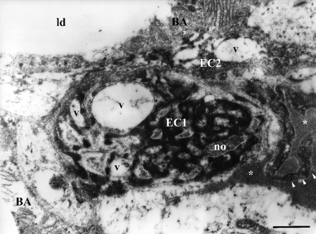
Figure 3.
Ultrastructure of apoptotic endothelial cells in brown adipose tissue of hyperinsulinaemic rat (chronic treatment). EC1, endothelial cell with condensed, sparsely distributed chromatic material and with numerous vacuoles (v); EC2, neighboring endothelial cell also shows signs of apoptotic changes and organelle vacuolization, with protrusions (arrowheads) into capillary lumen (asterisks); BA, brown adipocyte; no, nucleolus; ld, lipid droplet. Scale bar: 1 µm.

