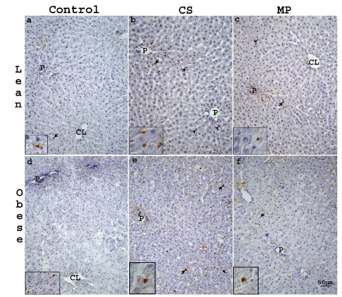
Figure 7.
Representative light photomicrographs of TUNEL-stained sections from liver of lean (a–c) and obese (d–f) Zucker rats. In control lean (a) and obese (d) livers only few hepa-tocytes (arrow heads) and SLC (arrows) were TUNEL positive. Both in lean and obese livers after CS (b,e) or MP20 (c, f) TUNEL-stained cells were located mainly in PP and MZ area. CL, centrolobular vein; P, portal vein. Scale bar: 50 µm.

