Fig. 2.
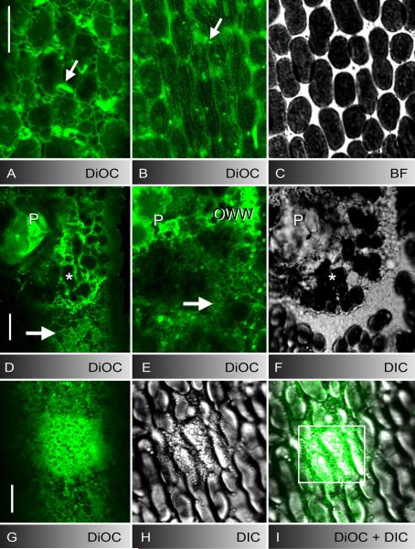
ER in control cells (A-C), at puncture (D-F) and UV-induced wounds (G-I). A-C DiOC6-stained cortical ER meshwork (A), subcortical ER tubes (B), and mitochondria (arrows) in unwounded cell regions and corresponding bright field image of cortical chloroplasts (C). D-F DiOC6-fluorescence (D and deeper optical section E) and DIC image (F) of a puncture wound. The wound plug (P) and cortical chloroplasts (asterisks) are surrounded and covered by brightly stained ER-remnants. The cortical ER in the unwounded control region (arrow in D) and the inner ER beneath the wound surface (arrow in E) is reticulate (compare with the tubular ER in control region B). G-I Accumulation of DiOC6-stained ER (G) and secretory vesicles (H) after 4 min UV-irradiation in the boxed area of the merged image I. Bars are 10 μm

