Fig. 4.
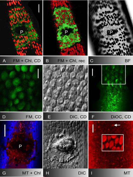
Actin-dependent deposition of FM1-43-stained membranes, secretory vesicles and ER cisternae at puncture (A-C) and UV-induced wounds (D-F) and behaviour of mitochondria (G-I). A-C FM1-43-stained membranes and autofluorescent chloroplasts in the outer wound wall one hour after puncturing in cytochalasin D (CD; A) and after two hours recovery in AFW (B; D is the corresponding bright field image). Note increase in FM1-43-fluorescence (A and B are 3-D projections of about 40 optical sections). D-F CD inhibits the migration and deposition of FM1-43-stained organelles (D), of secretory vesicles (E) and of DiOC6-stained ER cisternae (F) towards UV-induced wounds (images were taken after 5 min irradiation). Cortical ER is seen outside the irradiated area (boxed) in F. G-I Mitochondria labelled by mitotracker orange (red) are neither deposited at puncture wounds (autofluorescent chloroplasts are shown in blue; H is the corresponding DIC image) nor at UV-induced wounds (boxed area in I; damaged chloroplasts show enhanced autofluorescence). The arrow points to a cortical mitochondrion outside the irradiated area. P = wound plug. Bars are 10 (D-F and I) and 25 μm (A-C, G and H)

