Figure 4.
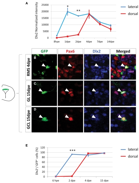
Dlx2 gets activated in the dorsal lineage once cells have entered the RMS and is maintained in the OB. (A) Evolution of Dlx2 transcript expression over time in the dorsal (red) and lateral (blue) lineages obtained from microarray analysis performed on FAC-sorted GFP electroporated cells at successive time points. (B–D) High magnifications of dorsally generated GFP+ cells labeled with Pax6 (red) and Dlx2 (blue) antibodies in the RMS at 4 dpe (B), or in the granule cell layer (C) and glomerular layer (D) at 15 dpe. Arrowheads: GFP+/marker+ cells. Note that both in the RMS and the GCL and GL of the OB Dlx2 is co-expressed with Pax6 in dorsally generated GFP electroporated cells. (E) Evolution of Dlx2 expression in dorsal (red) or lateral (blue) GFP+ electroporated cells at successive time after electroporation (n = 3 mice per group). Scale bar: 10 μm.

