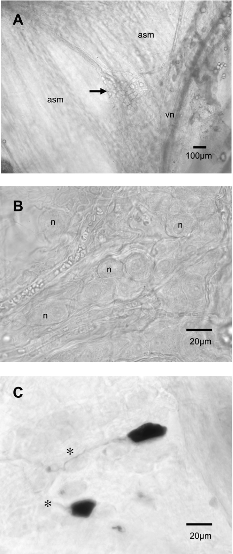
Fig. 1.
Micrograph images of airway parasympathetic ganglia. All neurons recorded were localized to ganglia near the tracheal bifurcation or on the adjacent right bronchus, near the vagus nerve. In A, a low-magnification view of a cluster of parasympathetic neuronal cell bodies (ganglion, arrow) found on the dorsal surface of the trachea on the airway smooth muscle (asm) near the vagus nerve (vn) in a fresh whole mount trachea (original magnification, ×4). In B, at higher magnification (×40), another ganglion located on the right bronchus. The somata of ganglionic neurons (n) were ovoid in shape. In C, after injection with Neurobiotin and having been fixed and developed using peroxidase and DAB, the neurons were ovoid to cuboidal in shape, lacked significant dendritic processes, and gave rise to a single axonal process (*); original magnification, ×40.

