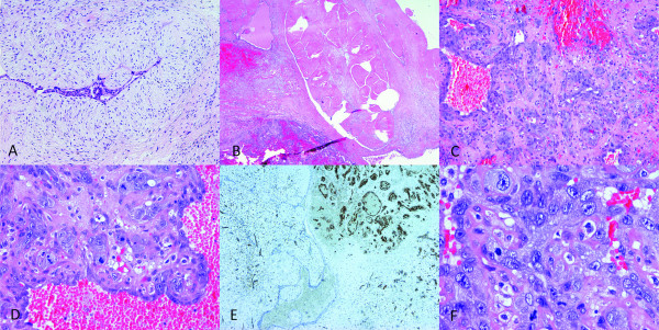
Figure 2.
Microscopic features of the resected tumor. Fibroadenoma of the breast HE, 100 × (A). Fibroadenoma and angiosarcoma in the same field, HE, 20 × (B). Angiosarcoma, the tumor is highly vascular with relatively solid spindle cell proliferation and area of stromal hemorrhage, HE, 100 × (C). Prominent tufts and papilations composed of enlarged highly malignant endothelial cells with brisk mitotic activity, HE, 200 × (D). Immunohistochemical stain for vascular marker CD34 was positive in angiosarcoma and negative in fibroadenoma, 40 × (E). Highly malignant endothelial cells, HE, 400 × (F).

