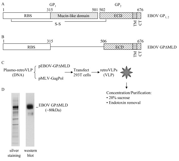
Figure 1.
Antigens used for immunization. A. Schematic diagram of the full-length GP1,2 of Ebola virus (EBOV GP1,2, GenBank# NC_002549) that encodes a polyprotein which upon cleavage yields two subunits, GP1 and GP2, linked together through a disulfide bond. GP1 contains the receptor binding site (RBS) and the highly variable, highly glycosylated, and dispensable mucin-like domain (MLD). GP2 contains an extracellular domain (ECD), a transmembrane domain (TM) and a cytoplasmic tail (CT). The numbers above the diagram represent the amino acid residue numbers. B. Schematic diagram of the resulting protein expressed by plasmids used for immunization studies or to derive in vitro VLPs. EBOV GPΔMLD was deleted in the MLD domain and the cleavage site between GP1 and GP2 [40], so the resulting protein expressed is a single molecule. C. Production scheme for VLPs. Supernatant from HEK 293T cells containing VLPs was centrifuged through a 20% sucrose cushion to concentrate and partially purify the VLP, which was further purified to remove endotoxin. D. Verification of EBOV GPΔMLD incorporation into VLPs. The VLP product was resolved through a NuPAGE 4-12% Bis-Tris gel, the total protein was evaluated with silver staining, and EBOV GPΔMLD was detected by western blot.

