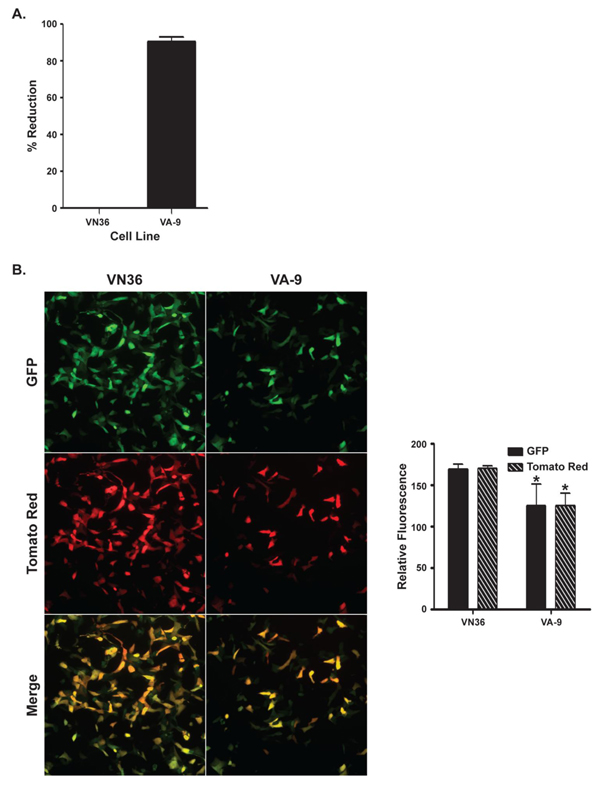
Figure 7.
Infection of cells constitutively expressing MxA. (a) VA-9 and VN36 cells were infected with MPXV-Zaire at an MOI = 5. The cells were harvested and lysed 24 h p.i., and the amount of virus present in the lysates was titered by plaque assay. (b) VA-9 and VN36 cells were infected with MPXV-GFP-tdTR at an MOI = 5. The cells were imaged 24 h p.i. by fluorescence microscopy (left), and relative fluorescence was measured using a fluorescence microplate reader (right). Green is GFP, red is Tomato Red, and yellow represents the overlap of green and red fluorescence.

