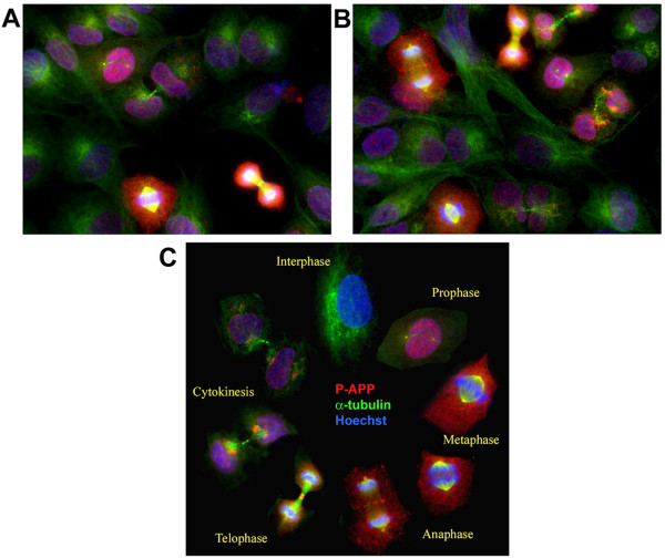
Figure 10.
Analysis of P-APP distribution in asynchronously growing H4-15X cells show cell cycle-dependent localization of P-APP: Panels A and B show asynchronously growing H4-15X cells fixed and immunostained using Thr668 P-APP polyclonal and α-tubulin monoclonal antibodies and visualized using Alexa 594 (red) and 488 (green) fluorophores respectively. Staining was analyzed using the AxioVision Rel 4.8 software for Zeiss microscope. Nuclei were visualized using Hoechst staining. Cells in mitotic phase showed P-APP localized to the centrosomes, nucleus and cytoplasm with maximum immunoreactivity in mitotic cells and minimum/none in interphase cells. Cells in telophase showed P-APP staining in the midbody which was absent in cells undergoing cytokinesis. The absence of staining in the interphase cells suggests that APP phosphorylation at Thr668 occurs only when cells are undergoing division. Panel C shows a cell cycle schematic with representative cells from different stages of the cell cycle (selected from an asynchronously growing culture) illustrating the phosphorylation event occurring once the cells enter prophase and tapering off as it exits the cell cycle (cytokinesis). Magnification: 63×.

