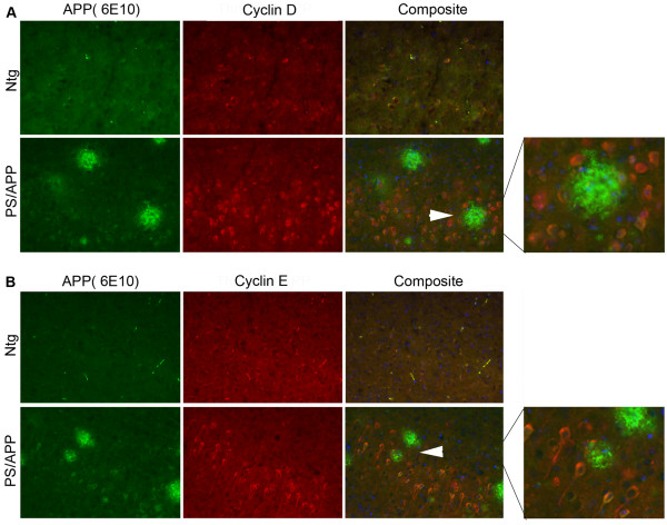
Figure 2.
AD transgenic mice show increased expression of cyclin D and cyclin E in neurons: Brain sections from 10 month old Ntg and PS/APP mice were co-stained using A) monoclonal 6E10 and polyclonal cyclin D1 or B) 6E10 and polyclonal cyclin E antibodies. Staining was visualized using Alexa fluor 488 (APP and Aβ, green) and Alexa fluor 594 (red) and analyzed under a Zeiss microscope using AxioVision Rel 4.8. The images were taken at 20× magnification. The composite image shows staining with Hoechst, cyclin, and 6E10 antibodies. The area indicated by arrows is enlarged and shown on the right to clearly see the positive staining in neurons.

