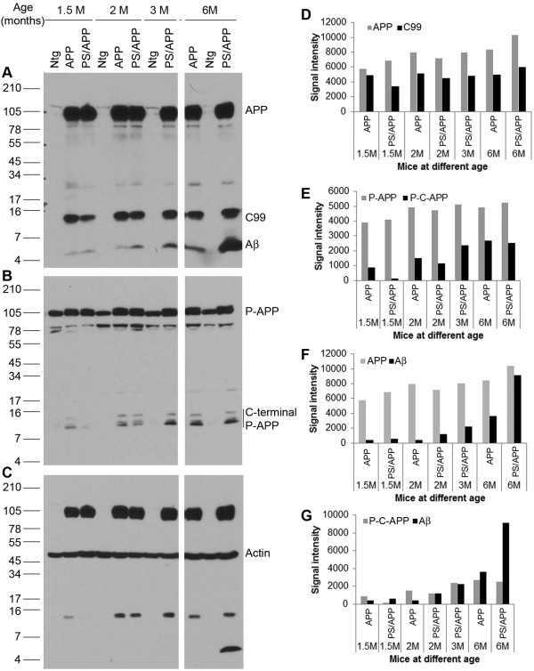
Figure 6.
Age-dependent changes in Thr668 specific phosphorylation and Aβ generation in transgenic mice: Brain extracts from 1.5, 2, 3, and 6 month old Ntg mice and transgenic mice expressing APP and PS/APP were examined by western blot using Thr668 P-APP and 6E10 antibodies. Panel A shows the levels of full length APP and fragments of APP such as C-99 and Aβ in the mice at different ages. The transgenic mice expressing APP and PS/APP showed very high levels of full length APP. Only the levels of Aβ were altered in an age-dependent manner. Panel B shows staining of the blot with Thr668 P-APP antibody, which detects mouse and human APP phosphorylated at this site. The levels of full length P-APP were higher in the transgenic mice. Levels of phosphorylated C-terminal P-APP fragments were induced in an age-dependent manner in the transgenic mice. Panel C shows reprobe of the blot with actin antibody without stripping to show approximately equal amount of protein loading. D-F shows the relative signal intensity of the various APP fragments from the western blot analysis. D) Represents signal intensity of full-length APP and C-99 fragments, E) that of full length P-APP and phosphorylated C-terminal fragments of P-APP (P-C-APP), F) represents the levels of APP and Aβ and G) shows signal intensity of P-C-APP and Aβ.

