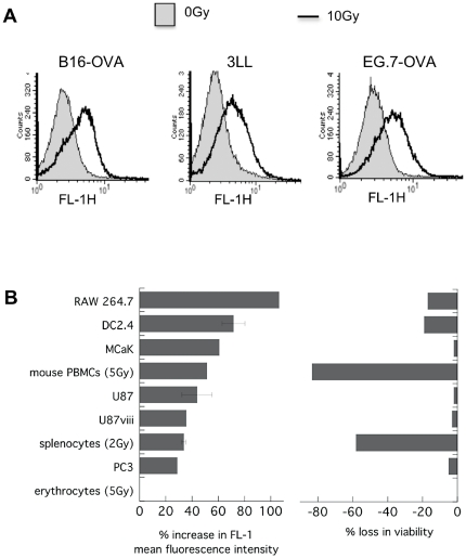
Figure 2. Many cell types respond to radiation with a rise in autofluorescence.
Various murine and human cell lines and primary mouse peripheral blood cells were irradiated with 10 Gy (or less when indicated) and left under standard culture conditions for 24 h prior to FACS analysis. Autofluorescence was recorded in FL-1 and cell viability assessed according to 7-AAD dye exclusion in FL-3. A) FL-1 histogram overlays for treated and untreated B16-OVA, 3LL and EG.7-OVA cells. B) Percent increase in mean FL-1 fluorescence and percent loss in viability in the irradiated sample as compared to control of n = 1–8±s.e.m.

