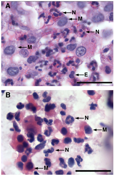
Figure 4. Pulmonary inflammatory response to antigen challenge in OVA-sensitized rabbits.
Representative high magnification photomicrographs (mag. ×1000) demonstrating that inflammatory infiltrate in lung tissues (A) and BALF (B) isolated from OVA-sensitized+challenged rabbits is mostly composed of neutrophils (N) with lesser amounts of mononuclear macrophages (M) and rare eosinophils (E).

