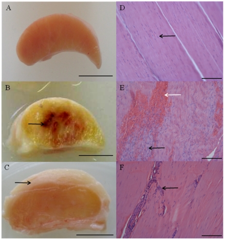
Figure 1. Typical macroscopic appearance of normal and injured equine flexor tendons.
(A) Normal superficial digital flexor tendon (SDFT) from a 12 year old horse; (B) sub-acutely injured SDFT 3 weeks post injury from a 4 year old horse, exhibiting a haemorrhagic granular central core (arrow) and (C) chronically injured SDFT >3 months post injury with a thickened fibrosed paratenon (arrow) from a 12 year old horse. Scale bar for macroscopic images = 1 cm. Corresponding longitudinal histology sections stained with Haematoxylin and Eosin: (D) normal SDFT showing regular arrangement of collagen fibrils (arrow); (E) sub-acutely injured SDFT with marked cellular infiltration (black arrow) and haemorrhage (white arrow); (F) chronically injured SDFT with increased cellularity in peri-vascular regions (arrow). Histology scale bar = 12.5 µm.

