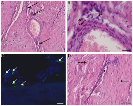
Figure 2. Haematoxylin and Eosin stained longitudinal histology sections of chronic injured SDFT (>3 months post injury) from a 7 year old horse (A, B and D).
(A) Reactive fibroplasia with increased cellularity in peri-vascular region (arrows). Scale bar = 100 µm. (B) Higher magnification of (A) showing presence of macrophages (arrowheads) in peri-vascular areas. Scale bar = 20 µm. (C) Corresponding 3-dimensional reconstructed Z stack image of dual antibody labelling for CD14 (red) and CD206 (green). Blue represents Hoechst nuclear counter stain. White arrows show CD14lowCD206high M2Mϕ located in peri-vascular endotenon regions. Scale bar = 20 µm. (D) Histological appearance of more normal SDFT to the right of the image (straight arrow) in contrast to irregular arrangement of collagen fibrils on the left (dashed arrow). The linear interface between more normal and injured zones of tendon is demarcated by an area of increased cellularity containing macrophages (arrowhead). Scalebar = 50 µm.

