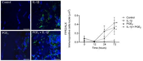
Figure 9. Panel of representative 2-dimensional images illustrating FPR2/ALX expression in tendon explants showing non-stimulated (vehicle only) control compared to explants stimulated with 5 ng ml−1 IL-1β and 1.0 µM PGE2 either alone or in combination.
Immunopositive staining is green, with Hoechst nuclear counter stain in blue. Scale bar = 12 µm. Graph showing the effect of pro-inflammatory mediators on tendon FPR2/ALX expression in vitro. Data represent average FPR2/ALX expression in tendon explants derived from 3 normal (uninjured) horses, whereby 2 replicates were analysed for each experimental condition and time point per horse. FPR2/ALX expression was determined at time points 0, 12, 24 and 72 hours after stimulation and compared to non-stimulated controls. Significant up-regulation of FPR2/ALX occurs 24 hours after stimulation with IL-1β or a combination of both mediators, and maximally 72 hours after stimulation with IL-1β, PGE2 or combination of both mediators compared to controls. ** P<0.01, * P<0.05. Error bars denote SEM.

