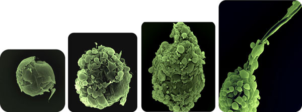
Figure 1.
Microfollicle formation. SEM images taken from developing microfollicles. After adding keratinocytes and melanocytes to the culture medium a loosen attachment to the neopapilla is seen (A). Polarization of the early aggregate (B). Assembly, orientation and sheath formation (C); microfollicle with fiber production (D).

