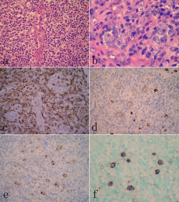
Figure 1.
Histopathological findings of lymph node at the initial presentation. (A) The architecture of lymph node was effaced by a diffuse polymorphic infiltration composed of small- to medium-sized lymphoid cells, immunoblastic cells and scattered eosinophils around high-endothelial venules. (B) Multinucleated cells with eosinophilic nucleoli resembling Reed-Stemberg (RS) cells could be observed in the lymph node. (C) Immunohistochemical examination revealed that infiltrated small to medium-sized lymphoid cells were diffusely positive for CD3, but large immunoblast-like cells and RS-like cells were positive for CD20 (D) and CD30 (E).These larger cells were also positive for EBER by in situ hybridization (F).(A, H&E staining, with original magnification ×400; B, H&E staining, with original magnification ×600; C-E, immunohistochemical staining, with original magnification ×400; F, EBER-in situ hybridization, with original magnification ×400).

