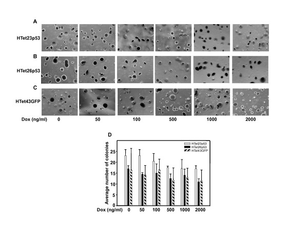
Figure 3.
p53 over expression did not reduce colony number. Five thousand cells were plated in low-melting agarose containing complete medium and allowed to grow for 30 days. (A, B and C) Cells were stained with crystal violet and photographs were taken under microscope for HTet23p53, HTet26p53 and HTet43GFP plates. (D) Colonies were counted and average number of colonies was plotted vs Dox concentration. Bar represents average of colonies from two representative fields (± SE).

