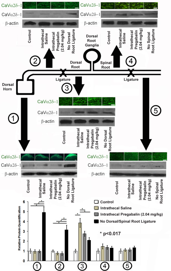
Figure 7.

Dorsal and Spinal Root Ligation Experiments. A cartoon diagram of the procedures performed in spinal nerve ligation and dorsal root ligation is provided. Tissues obtained after 7 days of spinal nerve ligation and/or dorsal root ligation (or neither in control rats) were examined using immunohistochemistry for CaVα2δ-1 protein and with Western blotting for CaVα2δ-1 protein and β-actin, used as a loading control. At the level of the dorsal horn (1), the presence of a dorsal root ligature prevented upregulation of CaVα2δ-1 protein regardless of delivery of intrathecal pregabalin or intrathecal saline. At the dorsal root proximal to dorsal root ligature (2), there was a similar lack of upregulation of CaVα2δ-1 protein regardless of delivery of intrathecal pregabalin or intrathecal saline, unless dorsal root ligature was absent. At the level of the dorsal root distal to spinal nerve ligation (3), there was an accumulation of CaVα2δ-1 protein, with less significant CaVα2δ-1 protein expression when intrathecal pregabalin was delivered or if spinal nerve ligation was absent. Distal to the dorsal root ganglia and proximal to spinal nerve ligation (4) or at the spinal root distal to spinal nerve ligation (5), there was no change in expression of CaVα2δ-1 protein irregardless of delivery of intrathecal pregabalin or intrathecal saline, or even if spinal nerve ligation was absent. Multiple unmatched ANOVA tests were performed between cohorts receiving intrathecal pregabalin and saline as compared to control rat samples and samples without placement of either spinal or dorsal root ligatures, with * indicating significant difference (p < 0.017 after Bonferroni corrections) between cohorts. Bars = 10 μm.
